About HKJR
Hong Kong Journal of Radiology (HKJR) is the official peer-reviewed academic journal of the Hong Kong College of Radiologists. HKJR is published quarterly by Hong Kong Academy of Medicine Press. HKJR is a continuation of the Journal of the Hong Kong College of Radiologists.
HKJR publishes papers on all aspects of diagnostic imaging, clinical oncology, and nuclear medicine, including original research articles, review articles, perspectives, pictorial essays, case reports, brief communications, editorials, and letters to the Editor. Papers on radiological protection, quality assurance, audit in radiology, and matters related to radiological training or education are also included.
The 2024 Journal Impact Factor for the HKJR is 0.2 (Clarivate, 2025).
FREE full text of ALL issues is available.
Additional materials may be made free at the Editorial Board's discretion.
Online First articles
Online First articles are released before they are included in a journal issue. These articles are fully citable and come with a DOI, enabling the most recent research to be accessed promptly.
Current Issue
Volume 29 Number 1, March 2026
![]() FULL TABLE OF CONTENTS Download the full issue
FULL TABLE OF CONTENTS Download the full issue
Highlights of this issue
About the Cover Images
 |
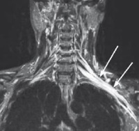 |
| In the article “Perineural and Muscular Involvement in Recurrent Diffuse Large B-Cell Lymphoma Detected by Fluorine-18 Fluorodeoxyglucose Positron Emission Tomography/Computed Tomography: A Case Report”. Recurrence and suspected atypical lymphomatous involvement in neuromuscular regions. Fused positron emission tomography/computed tomography image shows a hypermetabolic right neck neuromuscular lesion over the right trapezius muscle and accessory nerve region (arrow). | In the article “Revisiting Preoperative Evaluation of the Inferior Vena Cava in Abdominal Malignancies: A Pictorial Essay”. Posterior volume-rendered contrast enhanced computed tomography image shows a left inferior vena cava with hemiazygos continuation, crossing the midline posterior to the aorta (A) and draining into the azygos-superior vena cava pathway (white arrows). Hepatic veins are visible draining separately into the right atrium (green arrow). |